Skin infections, such as cellulitis, can sometimes be difficult to distinguish from other inflammatory conditions. Imaging modalities like ultrasound can help identify the presence of fluid collections or abscesses, confirming the diagnosis of a skin infection. This is particularly useful in cases where clinical examination alone is insufficient.
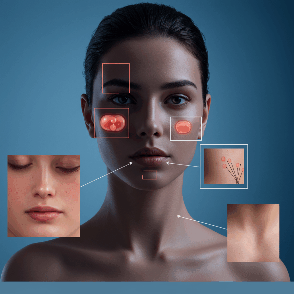
Rosacea:
Rosacea is a chronic skin condition with symptoms like facial flushing and raised bumps. While typically diagnosed clinically, imaging can help rule out other conditions with similar presentations. Conditions like lupus or certain types of acne can mimic rosacea, and imaging can help differentiate between them. Imaging can also be used to assess the severity of rosacea and guide treatment decisions.
Actinic Keratosis:
Actinic keratosis appears on body parts that are exposed to the sun, which include hands, arms, face, scalp, and neck. The sizes are normally less than two cm or nearly the size of an eraser. They are thick, scaly, or like crusty skin patches, typically pink in color with a brown, tan, or gray base. Imaging techniques can highlight abnormal skin structures. If left untreated, actinic keratosis can develop into squamous cell carcinoma, making early detection and treatment crucial.
Melasma:
Melasma presents as dark patches on the face. Melasma is very common in pregnant women and those with darker skin color or individuals greatly exposed to the sun. Imaging can assist in differentiating it from other pigmentary disorders. Different types of melasma exist, and imaging can help differentiate between them, guiding treatment strategies.
Warts:
Warts, caused by HPV or human papillomavirus, can be visualized using imaging to determine their extent and guide treatment. It could appear on the skin or in mucous membranes and might occur singly or in groups. It is important to note that Wart is infectious and could infect others. Early detection and treatment of warts are crucial to prevent their spread.
Hives:
Hives occur after exposure to an allergen and have itchy, raised welts that are red in color, warm, and mildly painful when touched. Imaging can help in complex cases. In complex cases, imaging can help rule out other conditions that may present with similar symptoms.
Conclusion:
Medical imaging is revolutionizing the diagnosis and management of various skin conditions. From early skin cancer detection to differentiating inflammatory dermatoses, these technologies offer valuable insights that improve patient care. Improving access to skin disease information is crucial. Furthermore, addressing the disparities in medical imaging for darker skin tones is essential for equitable healthcare.